Grade 1 Left Ventricular Diastolic Dysfunction With Normal La Filling Pressure
Normal mitral valve anatomy. The American Journal of Medicine - The Green Journal - publishes original clinical research of interest to physicians in internal medicine both in academia and community-based practiceAJM is the official journal of the Alliance for Academic Internal Medicine a prestigious group comprising internal medicine department chairs at more than 125 medical schools across the US.
Diastolic Dysfunction Cardio Guide
INTRODUCTION Heart failure with preserved ejection fraction HFpEF is a clinical syndrome in which patients have symptoms and signs of HF as the result of high ventricular filling pressure despite normal or near normal left ventricular ejection fraction LVEF 50 percent Most patients with HFpEF also display normal LV volumes and evidence of diastolic dysfunction eg.
Grade 1 left ventricular diastolic dysfunction with normal la filling pressure. A fibro-muscular annulus two leaflets tendinous chords and papillary muscles In order to identify abnormal mitral anatomy by echocardiography and accurately diagnose. Anything that decreases right atrial pressure downward early systolic atrioventricular septal motion early diastolic right ventricular filling will cause the wave to slope downward. The normal mitral valve MV sits at the junction between the left atrium LA and left ventricle LV.
Figure 19 Normal time-correlated electrocardiographic ECG findings central venous pressure CVP tracing and hepatic venous HV waveform 4. An LVEF of 50 or or more with symptoms of heart failure is referred to as heart failure with preserved ejection fraction HFpEF formerly known as diastolic heart failurean indication that as a result of remodeling the heart muscle has become thick or rigid preventing the normal volume of blood from filling up ventricles relaxation. It is a complex anatomical structure composed of several distinct but contiguous structures.
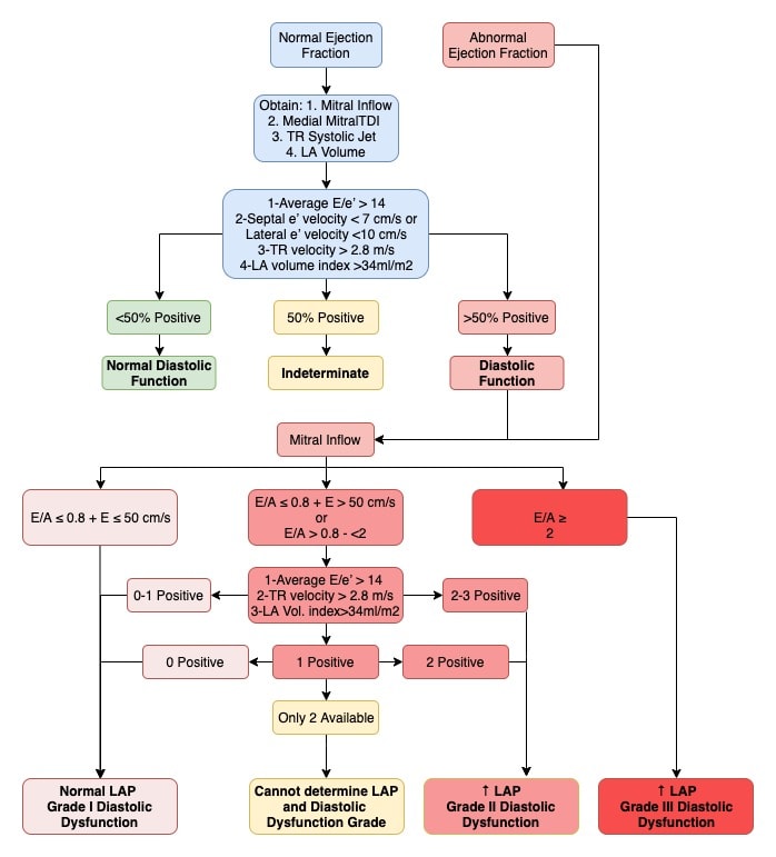
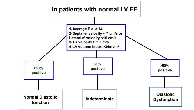
Normal LAP Grade I diastolic dysfunction 3 criteria to be evaluated 1 Average Ee14 2 TR velocity 28 ms 3 LA volume index 34 mLm2 2 of 3 negative 2 of 3 or 3 of 3 positive 2 negative 1 positive and 1 negative 2 positive When only 2 criteria are available Cannot determine LAP and diastolic dysfunction grade LAP Grade II.

Left Ventricular Diastolic Function Thoracic Key
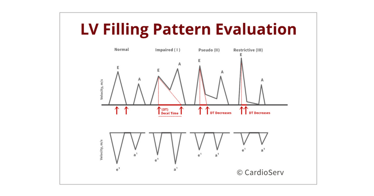
Understanding The Basics Lv Filling Patterns
Understanding The Basics Lv Filling Patterns

Left Lv Diastolic Pressures Recording Arrows Point To Lv Minimal Download Scientific Diagram

Pdf Left Ventricular Diastolic Function And Dysfunction Central Role Of Echocardiography Semantic Scholar

Key Predisposing Left Ventricular Diastolic Dysfunction Increased Download Scientific Diagram
![]()
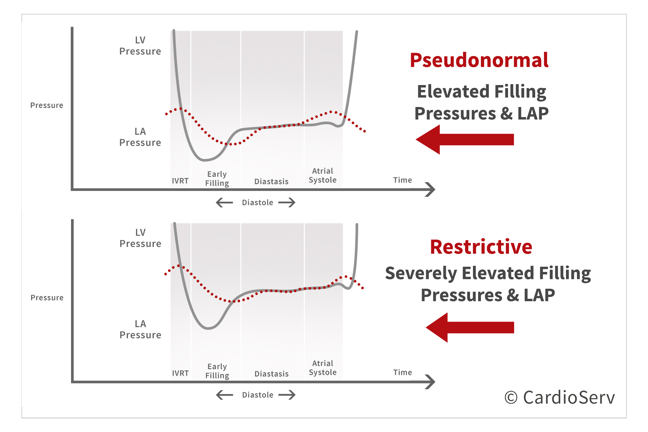
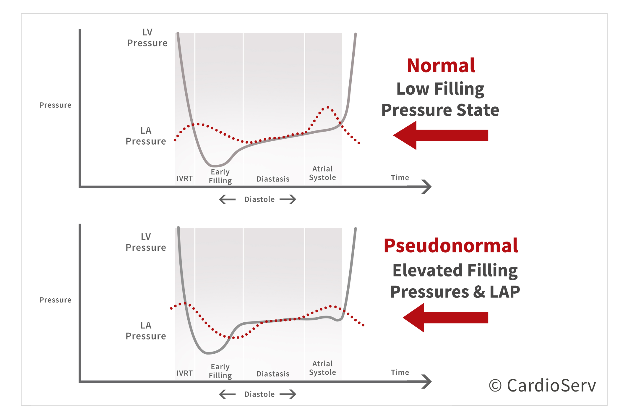
Left Ventricular Lv And Left Atrial La Pressures During Diastole Download Scientific Diagram

Grades Of Diastolic Dysfunction Download Table

The Stages Of Diastolic Heart Failure Lv And Left Atrial La Download Scientific Diagram

Pathophysiology And Echocardiographic Diagnosis Of Left Ventricular Diastolic Dysfunction Journal Of The American Society Of Echocardiography
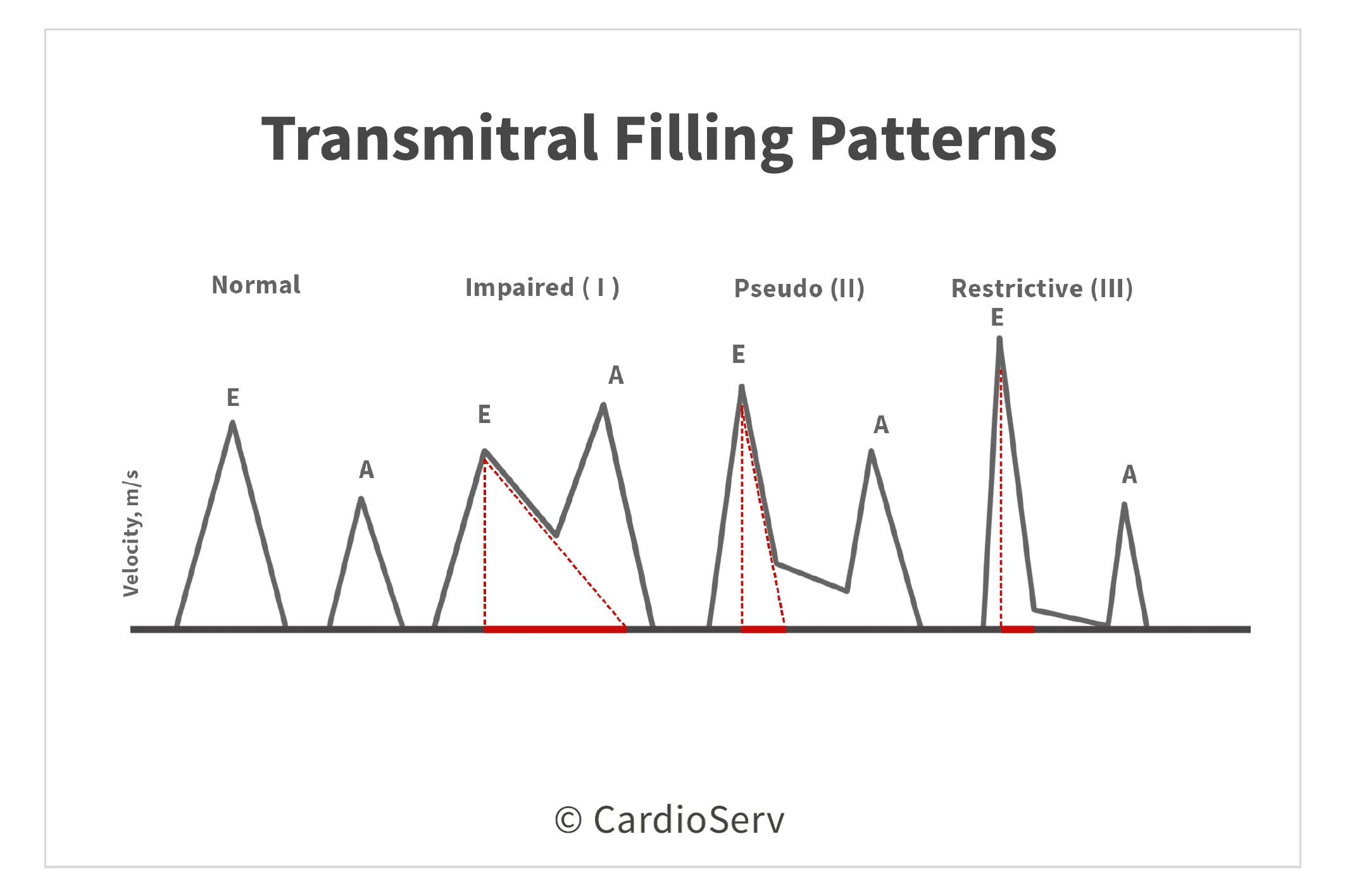
Understanding The Basics Lv Filling Patterns
4 5 A Simple Approach To Diastolic Dysfunction 123 Sonography

Diastolic Dysfunction Learn The Heart

Pathophysiology And Echocardiographic Diagnosis Of Left Ventricular Diastolic Dysfunction Journal Of The American Society Of Echocardiography
Understanding The Basics Lv Filling Patterns

Assessment Of Diastolic Function By Echocardiography Ecg Echo

Diastolic Dysfunction Nursing School Survival Pharmacology Nursing Cardiology Nursing

Practical Approach To Grade Diastolic Dysfunction By Echocardiography Download Scientific Diagram

Left Atrial Strain As A Single Parameter To Predict Left Ventricular Diastolic Dysfunction And Elevated Left Ventricular Filling Pressure In Patients Undergoing Off Pump Coronary Artery Bypass Grafting Journal Of Cardiothoracic And